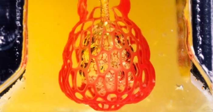
Scientists are continuously working to improve the technology of 3D printing when creating artificial organs for human transplantation. However, as a result of the creation of certain organs, there is a problem associated with a complex structure consisting of numerous small vessels that are densely intertwined (multivascularization).
Creating a copy of human organs from hundreds of intertwined capillaries and vessels for pumping blood, lymph and air was made possible due to the development of the scientists from the United States. The new technology of three-dimensional biological printing uses stereolithography apparatus for tissue engineering or SLATE. The 3D printer uses a hydrogel, which dries when exposed to ultraviolet light, and the organs are printed in layers with high resolution (10-50 microns).
The invention was tested to create an analog of human lungs that are strong enough and resilient to withstand repeated cyclical compression and expansion when air passes through them. In the future, it is planned to create not only organs intended for transplantation but also test samples, on which it will be possible to study the process of formation and development of cancer cells. In the meantime, the prototype is light in size – less than a one-cent coin. The task of the researchers is to achieve the scalability of the organ to the desired size without loss of functionality and endurance during continuous operation over the years.


